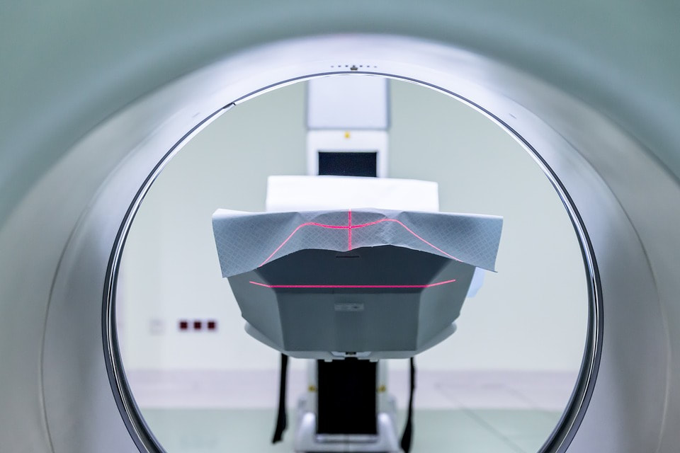
BRAND NEW STUDY SHOWS DIFFERENCES IN MRI IMAGING OF FASCIA IN AUTOIMMUNITY

“Autoimmune disorders of the musculoskeletal fascial system include numerous diseases such as systemic sclerosis, lupus, eosinophilic fasciitis, dermatomyositis, polymyalgia rheumatica and some other mixed connective tissue diseases along with seronegative and seropositive arthritides. Each disease tends to target specific components of the fascial system with inconstant involvement of the adjacent structures.
According to the most recent definition of the fascial system, fasciae also include identifiable anatomical structures (retinacula, interosseous membranes, tendons and entheses) and connective tissues specific to organs like muscles (epimysium and perimysium), nerves (epineurium and perineurium) and vessels (adventitia) MRI is the most efficient imaging technique to assess inflammatory changes of the fascial system.”From the study being discussed today
Almost a year ago to the day, I gave you the ever popular post, FASCIA AS RELATED TO AUTOIMMUNE DISEASES. I’ve also shown you over and over that contrary to the public has been led to believe, MRI does not do a great job of imaging soft tissues and won’t show the FASCIAL ADHESIONS I deal with in my clinic all day long.
However, if you are imaged with a newer unit with a larger magnet, current research is showing that it is possible to image diseased fascia in certain autoimmune diseases. A dozen French and Belgian researchers writing in the brand new issue of Insights Into Imaging (Fasciae of the Musculoskeletal System: Normal Anatomy and MR Patterns of Involvement in Autoimmune Diseases) put it this way…..
“The fascial system is a complex network of connective tissue that interconnects all the components of the musculoskeletal system. The normal fascial system is relatively inconspicuous at MRI. MR patterns of fascial involvement in autoimmune diseases reflects the complex anatomy of the musculoskeletal fascial system.
Autoimmune inflammatory disorders may involve the fasciae as well as the other components of the connective skeleton. There are however important variations among these conditions concerning the target tissue, intensity of the inflammation, type of cellular infiltrates, their natural history and, finally, their influence on the adjacent tissue such as the synovium, muscles or bones.”
So; what I’ve been telling you all along is true; IMAGING FASCIA is difficult at best. However, if a radiologist is good, they can spot slight variations of normal (lots of side by side images of normal compared to pathological in this free study). Allow me to give you an example. The PERIOSTEUM is the fascial membrane that covers bone. As you notice what these authors say about the periosteum as related to AUTOIMMUNE DISEASES, pay attention to the word “THICKEN” and remember that one of the characteristics of messed up fascia is that it thickens when things go south.
“MRI involvement of the periosteum in active autoimmune diseases consist of thickening…. Periosteum can be the main target as in hypertrophic [thickening] osteoarthropathy [bone arthritis] or accompany involvement of the deep fasciae or connective tissue of the muscles.“
The authors talked about the epi and perimysium as well (the fascial layers that surround and permeate muscles), saying that “On MRI, it may be impossible to distinguish involvement of the connective tissue [fascia] from involvement of the muscle fibres. These changes may be focal or diffuse and may involve whole muscles or muscle groups. Chronic inflammatory involvement may induce fatty transformation of the muscles.”
This last sentence is important because it was just a couple of days ago I mentioned “fatty infiltration” in another post (HERE). For the record, “thickening” was discussed in other fascia-involved tissues as well: RETINACULA, APONEUROSES, MUSCLES themselves, as well as TENDONS. As far as the superficial and deep fasciae are concerned, the authors said this….
“MR involvement of the superficial fascia in active autoimmune diseases consists of thickening of the hypodermic reticular network of the superficial fascia…. Involvement of the superficial fascia can extend deeply and involve the deep peripheral fascia and the connective tissue of the muscles.
MRI involvement of the deep fasciae in active autoimmune diseases consists of thickening of the deep peripheral fascia between hypodermis [skin] and muscles and/or the deep intermuscular fascia between muscles…. Deep fasciae are the main target of eosinophilic fasciitis and involvement can extend to the adjacent structures and involve superficial fascia, epi- and perimysium and/or periosteum. Chronic involvement of the deep fasciae may induce persistent thickening of the fasciae… suggesting fibrosis.”
There it is again — thickening. Only this time we see it linked to something I discuss at length on this site; FIBROSIS, which is essentially another name for scar tissue (HERE). All of this imaging stuff is great, but I’m not really sure that it’s very helpful. As always, the bottom line is whether or not there is anything to be done about it.
Because autoimmune diseases are ultimately problems caused by a runaway immune system that’s decided to attack self, it’s important to understand that the drugs used to treat these various diseases SUPPRESS THE IMMUNE SYSTEM (don’t even think about “BOOSTING” the immune system of a person with autoimmunity).
Because it’s all based on inflammation, if you can find what’s driving said inflammation, while you may not be able to “cure” your problem, in many cases you can dramatically slow it’s progression and improve your symptoms in the process. How? HERE. And since you probably know someone who is struggling with autoimmunity (HERE is a short list), be sure to share this post with them (FACEBOOK is a great way to do this).
