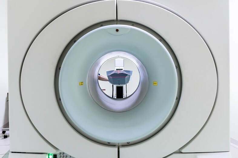
NEW STUDY TOUTS MERITS OF MRI IMAGING FOR MUSCULOSKELETAL PROBLEMS / PAIN

“Despite its presence everywhere throughout the body, the fascial system has received little attention in the imaging literature as it is regarded as a network of inert membranes barely involved in abnormal conditions. In a previous article, we detailed how MRI patterns of involvement of the fasciae in systemic autoimmune diseases reflect the fascial anatomy. Anyway, the fascial system may also be involved in localized disorders. Therefore, the current pictorial review aims to focus on traumatic disorders, infectious diseases, and neoplastic diseases involving the fasciae of the musculoskeletal system and their appearance at MRI.” The statement above comes from the abstract (word-for-word) of a study published in the latest issue of the European journal, Insights Into Imaging (Fasciae of the Musculoskeletal System: MRI Findings in Trauma, Infection and Neoplastic Diseases). For those who are interested, I wrote about the relationship between fascia and autoimmunity (FASCIA AS RELATED TO AUTOIMMUNITY and FASCIA AS AN ENDOCRINE ORGAN) as well as the fact that these types of issues can often be imaged with MRI technology (MRI IMAGING OF FASCIA IN AUTOIMMUNE DISEASES). Not to sound like I don’t care, but imaging autoimmune diseases is not the thrust of today’s post. I’m far more interested as to what the experts have to say about imaging fascia in MUSCULOSKELETAL PAIN SYNDROMES.
The first thing to realize is that yes; some fascial injuries are going to show up on MRI for the simple reason that some of them (FASCIA HERNIATIONS for instance) can actually be seen with the naked eye. I’ve seen a couple of these in 30 years of treating patients — it’s not someting I can help with. The second important factor is knowing where to look. When it comes to connective tissues (bones, tendons, fascia, LIGAMENTS), these authors state that injury occurs as sites of “interfaces between the different components of the fascial system“. For example, in TENDINOSIS, there is a always a weak point where the fascia that makes up the PERIOSTEUM (the cellophane-like fascial membrane that covers bones) meets the tendon itself (tendons are also a form of fascia and are involved in various forms of tendinosis and TENDINOPATHY).
When there is enough force to overcome the tissue’s tensile strength, whether that force is due to “repeated microtraumas (overuse), acute injuries, or a combination of both,” the end result is the same — microscopic tearing and subsequent MICROSCOPIC FASCIAL ADHESIONS. The tissue fibers become deranged on a cellular basis, ending up with more in common with a hairball than with well-combed hair (HERE, HERE or HERE). As I’ve shown you repeatedly, these fascial tears / injuries / micro-traumas can also occur in many different ways (HERE). And while the authors mentioned various sorts of muscle issues; admittedly, the kinds of problems that are going to show up on MRI would be labeled as “tears” rather than what we refer to as MUSCLE PULLS, which are typically injuries of the — you guessed it — fascial sheath surrounding the muscle (the EPIMYSIUM).
The authors went on to discuss and show images of several different sorts of tumors and disease processes that affect fascia of the muscle and skeletal system. The thing to remember, however, is that despite advances in MRI technology (much larger magnets, which create better clarity), you cannot expect to see much beyond the obvious — aka, gross pathology. This is significant because many of you reading this who have either had MRI (s) or are hoping to get one soon, think that since your pain is so bad, your problem will show up with crystal clarity. In fact, many people erroneously believe that after they finally get their MRI, the doctor is going to look at them sympathetically and apologetically say, “Am I ever sorry for doubting your pain Mrs. Jones. Your fascia is so adhesed it’s no wonder you are suffering. In fact, if your problem had been any worse, our machine would have melted down to a puddle of molten steel. Let’s go solve this for you.” Those who have been through this know what I’m talking about.
That’s because with most of these kinds of problems, the underlying issues behind the chronic pain syndromes are functional and not pathological, meaning they don’t show up on any tests, MRI included (HERE). This is why so many of you who have been riding the MEDICAL MERRY-GO-ROUND have become all too familiar with the BIG FIVE CLASS OF DRUGS prescribed for chronic pain. It’s also why I urge patients who are going for MRI to temper their expectations (HERE).
So, despite the fact that other problems such as hernias (HIATAL or INGUINAL) or issues on the PALMAR APONEUROSES or plantar aponeuroses (usually referred to as the plantar fascia) can be seen with this technology, MRI imaging of musculoskeletal problems is almost exactly where it was when I wrote my 2014 post titled IMAGING THE BODY’S FASCIA. Since there’s little headway on the MRI front, this begs the question of what can be used to image problems of the fascia?
Certain types of fascial issues can be seen with diagnostic ultrasound (HERE), usually confined to tendons, the THORACOLUMBAR FASCIA (lower back) or PLANTAR FASCIA (bottom of foot). Having recently undergone a dynamic shoulder ultrasound with a brand new unit and a radiologist running it (I was curious), I can assure you that while certainly a move in the right direction, it will be awhile before this technology can do what the general public is hoping it can do, if it ever gets there. Fortunately, we can often see torn fascias and the subsequent FASCIAL ADHESIONS when examining and treating our patients (HERE, HERE or HERE are examples). Unfortunately, the paper concluded with these words.
“The musculoskeletal fascial system can be affected by various localized disorders with variable time course and prognosis. MRI is the best imaging technique to detect the presence of fascial lesions and assess their localization and extent, but it is limited for lesion characterization.”
“Limited“. Not a very comforting word if you struggle with CHRONIC PAIN. Allow me, however, to share with you a story from one of my patients who is too shy to do A VIDEO TESTIMONIAL for us. This person, who just graduated from professional school (congrats, BTW), was working out as a high school underclassman, when a heavy piece of steel weightlifting equipment (400 lbs or so) was pulled over and struck the individual squarely in the mid back. This person struggled for two years, seeing a number of specialists and undergoing several MRI’s. The results were the same, with this person finally being told outright that they were likely malingering (faking).
With the school’s liability insurance off the hook (a phenomenon that frequently occurs in WHIPLASH INJURIES as well), the family was now on their own. The first thing they did was come see me. After the very first treatment, this individual got up off the table and could not reproduce the pain — something that’s actually quite common in my clinic (HERE). What’s doubly interesting is that I showed the young patient’s mom the impact zone, who then validated it with photos. I see this individual once every year or two for a tissue remodeling treatment and adjustment.
The moral to the story is that whether you are dealing with CHRONIC NECK PAIN, HEADACHES, CHRONIC BACK PAIN, SHOULDER PAIN, or any number of others, there is likely to be a significant fascial component that is not showing up on the tests your doctors are running (HERE). This means it’s unlikely that it’s being addressed in any type of constructive manner. It also means that your problem is probably being blamed on age, DEGENERATION, DISC HERNIATION or one of the myriad of problems that readily show up on tests, but often have little clinical relevance (HERE and HERE are examples pertaining to the shoulder). It also means that it might be worth a visit to see.
If you are looking for solutions to these and other health-related problems, be sure to look at THIS SHORT SOLVE-IT-YOURSELF POST. And as always, if you appreciate what you are seeing, don’t forget to reach out to the people you love and value most by liking, sharing or following on FACEBOOK.

One Response
It is interesting that what manual therapists can actually palpate with their fingers, cannot be “detected” on sophisticated hi-tech imaging systems. What is the underlying problem here? Can some imaging system not produce an image of the “shape of the fascia sheet” anywhere? If a manual therapist can feel a knot in it, why can this not be “imaged”? Presumably it would detect a cancerous growth, so why doesn’t it detect a deviation in the “shape” of a healthy sheet of fascia?
One of the most infuriating arguments I have experienced from an “expert” entrenched in the “central nervous system” hypothesis, is that manual experts feeling and palpating something anomalous with their fingers is “not science” and should not be used as the basis for diagnosing anything. If sophisticated high-tech imaging systems “contradict” the existence of an anomaly that can be felt with probing fingertips, then I call bullshit on the imaging systems; their designers and operators don’t WANT to image the anomaly. Telling us it ain’t even there, is gaslighting us, and worse. It is psychological abuse, and the mental instability of some pain sufferers has probably been caused more by this kind of handling by medical experts, than even the pain itself.