TISSUE DEFORMATION: THE BEST METHODS FOR REMODELING FIBROTIC CONNECTIVE TISSUES

The soft tissues that I chiefly deal with in my clinic are LIGAMENTS, TENDONS, and FASCIA. Even though muscles are not considered to be “Connective Tissue,” for our discussion they will be simply because most (certainly not all) muscle injuries are actually injuries to fascia (HERE). Also, this list could include APONEUROSES, which are sort of a cross between Fascia and Tendons, but I usually just list them under “Fascia”.
These tough fibrous Connective Tissues are largely made up of an EXTRACELLULAR MATRIX (ECM), COLLAGEN, Elastin (just like it sounds, an extremely elastic fiber that has the ability return to it’s original shape after being stretched or stressed), and FIBROBLASTS — the cells that essentially build new Connective Tissues (for the record, if what you are doing is not stimulating fibroblastic activity, it won’t work).
I’ve had Staffordshire Bull Terriers for over 25 years (WE ARE ON OUR THIRD). These dogs need lots of activity and roughhousing. It’s what they were bred for. Fail to work these dogs out intensely, and they will be unhealthy. Likewise, your muscles and connective tissues need to be worked out. In fact, without REGULAR MECHANICAL STRESS, connective tissues and muscles will fall into a state of disarray and slow (or maybe rapid) degeneration (HERE).
Although it might come from work (around here I treat lots of ranchers and loggers), thanks to today’s sedentary society, it might need to come in the form of EXERCISE, STRETCHING, YOGA, WEIGHTLIFTING (strength or resistance training of some sort) etc, etc, etc. Bottom line, if you are not regularly stressing your connective tissues in multiple ways (and for that matter BONES, which are a non-fibrous connective tissue), you are causing yourself future grief. But what happens when these tissues are stressed too much?
There are any number of ways that connective tissues and muscles can be injured (HERE), with repetitive injuries typically being worse (HERE). When tissue damage occurs, INFLAMMATION is released (not nearly as much with TENDONS). Inflammation is what gets the ball rolling in most healing processes. The problem is, soft tissues often heal with something called FIBROSIS — the medical name for SCAR TISSUE.
While this situation is normal on some level and how your body is designed to heal and repair itself, it can and often does lead to problems — especially if you are not living an ANTI-INFLAMMATORY LIFESTYLE. Much of this has to do with the fact that Scar Tissue / Fibrosis is different from normal tissue in almost every conceivable way — not to mention it does not show up well with standard imaging (HERE).
This means that if you don’t want to live a life of pain, you’ll have to address the Fibrosis. For many people, this is relatively simple. In fact, for the majority of people in the majority of situations, SIMPLE STRETCHES OR EXERCISES are enough to get the job done. For others, CHIROPRACTIC ADJUSTMENTS or THERAPY provide enough mechanical stress to work through the minimally-fibrotic tissue and restore normal ROM.
For some of you, however, it will require “breaking” the fibrotic tissues that are TETHERING & RESTRICTING your joint’s ability to move properly, in order to get out of pain and restore function. The word “break” implies a degree of INTENSITY typically not seen with other forms of therapy. What does recent scientific literature have to say about this aspect of tissue remodeling?
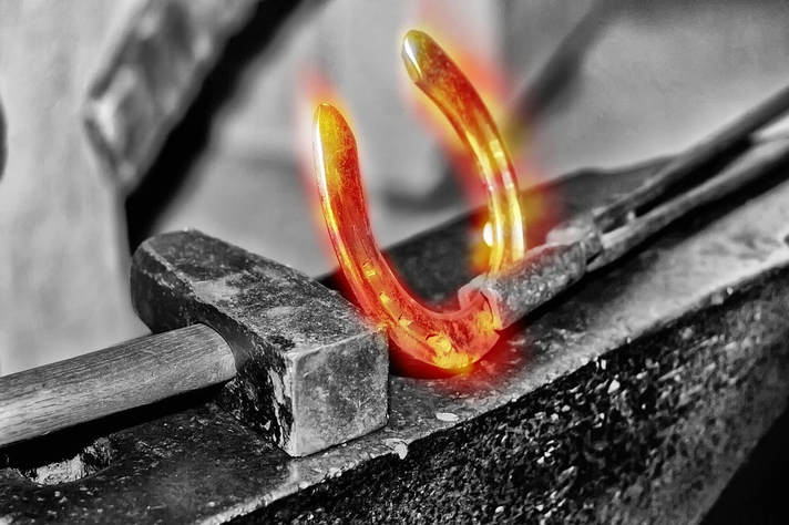
The first thing you must be aware of is that the scientific medical community usually refers to the purposeful mechanical stress put on tissues in order to remodel them as, ‘tissue deformation‘ (this term is also sometimes used to describe the injury process itself). In other words, if you want to make tangible, long-term changes to connective tissues and muscles, you must ‘deform’ them in some manner.
The second thing you need to be aware of is that there are dozens — maybe hundreds — of different mathematical and computer models of how this occurs. Unless you are really into advanced mathematics and computer algorithms, you won’t find it interesting.
I found “Tissue Deformation” studies about embryology, surgery (many of them pertaining to the development GM realistic-feeling tissues for surgeons to practice with), and even studies trying to figure out how much tissue deformation (i.e. sagging) the average female breast undergoes over time. Probably, however, the thing I was not expecting was that the huge majority of studies on this topic were theoretical. Allow me to explain.
Like I said just a moment ago, there are an insane number of computer and mathematical models being looked at concerning tissue deformation. Why is this? Why are we using models instead of real tissues, especially when according to 2012’s book, Virtual Reality in Medicine (chapter, Soft Tissue Deformation), “Mechanical tissue behavior is highly complex and only partly understood. Due to the complexity of soft tissue, the formulation of an appropriate mathematical model is a difficult task. Therefore, the accuracy of deformations can often only be roughly approximated.“?
Without going into incredible detail, suffice it to say that mechanical deformation of tissues is a far bigger factor not only in healing processes but in overall health than most of us have any idea.
This can be seen in the opening paragraph of a Dutch study from the Department of Tissue Regeneration, MIRA Institute for Biomedical Technology and Technical Medicine at the University of Twente. “Tissue deformation influences the development of the vasculature in the embryo and in the contracting wound. Current models suggest that physical forces originating from the blood, from cells pulling on neighboring cells, and on the Extra Cellular Matrix (ECM), distort cellular membrane receptors and cytoskeletal elements, modulating biochemical signaling pathways and the behavior of endothelial and smooth muscle cells.”
The cytoskeleton is made up of filaments, fibers, and tubules, and along with the ECM is made by FIBROBLASTS. Listen to what Wikipedia says about the cytoskeleton in relationship to tissue deformation.
“There is a multitude of functions that the cytoskeleton can perform. Primarily, it gives the cell its shape and mechanical resistance to deformation, and through association with extracellular connective tissue and other cells it stabilizes entire tissues. The cytoskeleton can also actively contract, thereby deforming the cell and the cell’s environment and allowing cells to migrate. Moreover, it is involved in many cell signaling pathways, in the uptake of extracellular material….. Furthermore, it forms specialized structures, such as flagella, cilia…..”
Tendons are also in on the act, with a study from last month’s issue of Acta Biomaterialia (Micro-Mechanical Properties of the Tendon-to-Bone Attachment) describing the forces between the velcro-like “Sharpey’s Fibers” that anchor tendons into the bones, and the bones themselves.
“The tendon-to-bone attachment (enthesis) is a complex hierarchical tissue that connects stiff bone to compliant tendon. The attachment site at the micrometer scale exhibits gradients in mineral content and collagen orientation, which likely act to minimize stress concentrations. To further examine structure-mechanical function relationships, local deformation behavior along the tendon-to-bone attachment was determined using local image correlation. A high compliance zone near the mineralized gradient of the attachment was clearly identified and highlighted the lack of correlation between mineral distribution and strain on the low-mineral end of the attachment. This compliant region is proposed to act as an energy absorbing component, limiting catastrophic failure within the tendon-to-bone attachment through higher local deformation.“
As you are probably beginning to see, tissue deformation can be either good or bad (remember that it describes both the injury and a mechanical portion of the process necessary for healing), and it can be painful. Last month’s issue of Clinical Biomechanics (Comparison of Lumbo-Pelvic Kinematics….)
“Compared to controls, individuals with acute low back pain had larger pelvic range of rotations and smaller lumbar range of flexions. Patients with acute low back pain also adopted a slower pace compared to asymptomatic controls which was reflected in smaller maximum values for angular velocity, deceleration and acceleration of lumbar flexion. Irrespective of participant group, smaller pelvic range of rotation and larger lumbar range of flexion were observed in younger vs. older participants.
Reduced lumbar range of flexion and slower task pace, observed in patients with acute low back pain, may be the result of a neuromuscular adaptation to reduce the forces and deformation in the lower back tissues and avoid pain aggravation.”
Certainly not surprising. Speaking of pain, how about pain in the face?
It is not uncommon for people who have CHRONIC NECK PAIN and / or HEADACHES, to end up with FACE PAIN. A study from the November 2016 issue of Skin Research and Technology (Analysis of Morphological Changes After Facial Massage…) looked at massages of the face using 3-D CT imaging, concluding that there were,
“marked morphological changes of the nasolabial folds after facial massage, and changes of the lower, upper and lateral cheeks and lower eyelid were also observed in more than half of the subjects. Facial massage-induced change rate values were significantly changed in the paranasal area, nasolabial fold area and cranial part of the mandibular area.
Photograph-based scores at the lower cheek and lower eyelid were well correlated with facial massage-induced change rate in the inferior part of the nasolabial fold and the mandibular area, respectively. Massage-induced changes of subcutaneous fat tissues and facial expression muscles were also apparent on CT images. 3D-CT imaging is useful for objective evaluation of the effects of facial massage, including anatomical changes in subcutaneous structures.”
The problem is that CT SCANS are highly toxic, as far as radiation exposure is concerned.
This information is probably helpful on some level, but for solving chronically adhesed fascia and other soft tissues, you’ll have to have more than information. Fortunately, I wrote a blog post concerning this issue, which helps explain why SIMPLE STRETCHES are often times not enough to solve these sorts of problems (as well as why it can sometimes actually make them worse).
To see my blog post on why it is necessary to cause TISSUE DEFORMATION (in this case for people with neck problems) just follow the link. For those of you dealing with whole-body issues, take a look at our PROTOCOL FOR ADDRESSING SYSTEMIC PAIN OR DISEASE. Once you begin to understand that most chronic conditions are variations of the same underlying screwed up physiological processes (HERE), it will all begin to make more sense. Be sure to like, share or follow on FACEBOOK if you appreciate the work I am doing here!
