
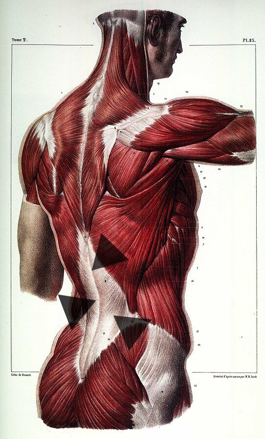
The THORACOLUMBAR FASCIA is arguably the single hottest area of study as far as low back pain is concerned. With LBP being almost ubiquitous in America (and increasing due to our sedentary, tech-driven lifestyles), it should pique your interest that the powers-that-be are now saying that almost 9 of 10 cases of low back pain (that’s 90% for those keeping score at home) are “non-specific” (HERE, HERE and HERE). Non-specific; huh? Listen to definitions of non-specific back pain as provided by experts.
Quotes From Experts on Non-Specific Back Pain (Thoracolumbar Fascia?)
The lifetime prevalence of low back pain in industrial countries is at 84%. Approximately 85% of such back pain is classified as non-specific, which means that no structural change, no inflammation and no specific disease can be found as its cause. The number of people who require treatment for back pain is high. From a 2019 issue of Frontiers in Psychology (Non-specific Low Back Pain and Postural Control During Quiet Standing—A Systematic Review)
Non-specific low back pain affects people of all ages and is a leading contributor to disease burden worldwide. Management guidelines endorse triage to identify the rare cases of low back pain that are caused by medically serious pathology, and so require diagnostic work-up or specialist referral, or both. Non-specific low back pain does not have a known patho-anatomical cause… Management consists of education and reassurance, analgesic medicines, non-pharmacological therapies, and timely review. The overuse of imaging, opioids, and surgery remains a widespread problem. From a 2017 issue of Lancet (Non-Specific Low Back Pain)
Non-specific low back pain has become a major public health problem worldwide. Mechanical factors, such as lifting and carrying, probably do not have a major pathogenic role…. History taking and clinical examination are included in most diagnostic guidelines, but the use of clinical imaging for diagnosis should be restricted. From a 2012 issue of Lancet (Non-Specific Low Back Pain)
Low back pain is defined as pain localised between the 12th rib and the inferior gluteal folds, with or without leg pain. Most cases are non-specific, but in about 10% of cases a specific cause is identified. LBP has a lifetime prevalence of 60–85%. At any one time, about 15% of adults have LBP. LBP poses an economic burden to society, mainly in terms of the large number of work days lost (indirect costs) and less so by direct treatment costs. From a 2007 issue of Best Practice & Research Clinical Rheumatology (Low Back Pain: Non-Specific)
Low back pain is a leading cause of disability. It occurs in similar proportions in all cultures, interferes with quality of life and work performance, and is the most common reason for medical consultations. Few cases of back pain are due to specific causes; most cases are non-specific. From a 2003 issue of the Bulletin of the World Health Organization (Low Back Pain)
Non-specific low back pain is one of the major reasons for medical consultations. As a definite somatic cause is identified in only 10% to 20% of cases. From a 1999 study in the British Journal of General Practice (Is Chronic Non-specific Low Back Pain Chronic? Definitions of a Problem and Problems of a Definition)
What do these definitions mean for you, the person trying to cope with chronic back pain? It means that despite one heck of a lot of time spent on the MMM, you have likely been dealt with in a manner not consistent with the info above —- lots of testing and drugs that are not really warranted, along with the possibility of lots of ineffective mechanical treatment (therapy, chiropractic adjustments, massage, acupuncture, etc). While these treatments are very frequently fabulous, they do not do a good job of addressing hardcore SCAR TISSUE so common in those with chronic, unremitting non-specific back pain (more on this later).
For a moment, however, let’s look at what the research medical community constitutes as “specific” low back pain.
Low back pain is a considerable health problem in all developed countries and is commonly treated in primary healthcare settings. It is usually defined as pain, muscle tension… with or without leg pain (sciatica). The most important symptoms of low back pain are pain and disability. About 90% of all patients with low back pain will have non-specific low back pain, which, in essence, is a diagnosis based on exclusion of specific pathology (radipulopathy, disc herniation, spinal stenosis, spondylolisthesis, ankylosing spondylitis, osteoporosis, lumbar spine fracture, skeletal mets [cancer], cauda equina syndrome, Scheurmans Disease, and scoliosis). From Physiopedia’s article, Specific Low Back Pain
Pay close attention to what these authors are saying.
Lots of people have low back pain — lots. Maybe even you. You’ve tried various things and it is not getting better. The insurance company finally authorizes your doctor to allow you to have some imaging done (MRI, CT, or maybe an old-fashioned x-ray). You are thinking to yourself — hoping — praying — that finally someone will ‘see’ why you are so miserable.
The problem is that many of you have this erroneous belief (KIND OF LIKE RALPHIE DID) that once the doctor ‘sees’ how terrible your back really is, he’ll collapse to his knees, sobbing and slobberingly apologizing for ever having doubted or underestimated your pain.
Stop! Wake up! Snap out of it! This is not reality for most of you.
What these authors are saying is that the mere presence of the problems listed above does not mean they are causing your pain and dysfunction. In fact, it is highly unlikely with most of them, particularly the most common of them such as disc herniations (the majority of which are “ASYMPTOMATIC“), OSTEOPOROSIS and DEGENERATION, which all have less to do with your pain than you have been led to believe. Probably way less.
(CANCER OF THE SPINE and CAUDA EQUINA SYNDROME, while rare, are not only dangerous, but crippling or even deadly. They are not common causes of back pain, however.)
With the medical community in agreement about the extent of non-specific low back pain, the question arises as to where said pain might be coming from. And maybe just as important, how might we know?
Although I have shown you several studies on imaging fascia (HERE, HERE, HERE and HERE), including THIS amazing ten-second video that I will suggest several times today to go back and watch, the fact remains that at least as far as “standard” imaging techniques are concerned, FASCIA does not show up well. Is there any hope on the horizon that this may be changing? Might we soon be imaging the thoracolumbar fascia on a regular basis?
Imaging the Thoracolumbar Fascia: Possibility or Pipe-Dream?
Since it’s the most commonly used advanced imaging modality, let’s look first at MRI as it pertains to our ability to visualize what’s going on with the thoracolumbar fascia.
After looking at the conclusions of a study published in September’s issue of the Asian Spine Journal (Evaluation of Diagnostic Accuracy of Magnetic Resonance Imaging in Posterior Ligamentum Complex Injury of Thoracolumbar Spine) I was hit with a dose of reality as far as MRI is concerned. In this fascinating study, the researchers took younger adults who had their thoracolumbar fascia seriously injured in car crashes, falls, etc, etc and were getting ready to undergo surgery.
The surgeons then compared the actual “injury” of the various soft tissues that make up the thoracolumbar fascia to their patient’s MRI, determining that an accurate diagnosis was made from magnetic resonance in just over a third of the cases. But here’s why that probably does nothing for you.
These were, as I said, people who were severely injured — injured to the point that the radiologists and surgeons were looking for and finding either partial or total disruptions of the tissues (LIGAMENTS, TENDONS, and APONEUROSIS) that make up the thoracolumbar fascia. Very few of you reading this have ever been in that boat. And even if you were at one time, that boat (surgical repair) has already sailed.
The point here is NOT TO BE AT ALL SURPRISED when your MRI does not provide the ‘ah-ha’ moment you were counting on / praying for. In fact, the authors concluded that the chances of an MRI diagnosing injuries to the thoracolumbar fascia considered to be the worst of the worst were even “lower than those previously reported in the literature.” Now; let’s shift gears, and as we do, provide a ray of hope. Let’s take a look at ultrasound technology as it pertains to imaging the thoracolumbar fascia.
Imaging the Thoracolumbar Fascia with Diagnostic Ultrasound
We’ll start the ultrasound discussion by mentioning a short paper from the April 2020 issue of the Journal of Ultrasound Medicine (Ultrasound Imaging of the Fascial Layers: You See Only What You Know). The authors, including renowned fascia researcher, Dr. Carla Stecco, made the point that ultrasound technology should be taking its rightful place as far as imaging soft tissues is concerned. In fact, these authors suggested that doctors can actually see far more fascia with diagnostic ultrasound than they are reporting in their radiology practices. Why is this?
Experts (both the people doing the test as well as those actually reading and interpreting the results) are not being trained to recognize fascial anatomy because it’s complex and time-consuming. The results is predictable — you do not “see” things you have not been trained to “see”. Everyone understands this. There is no one that cannot recite the old cliche, out of sight, out of mind.
Unfortunately, the authors determined that because “they are deemed unimportant, fasciae are likely to be underrated in the recent anatomy literature…. we strongly believe that ‘we conditionally see (only) what we know.’” Be sure to view the rest of today’s studies through this lens. In other words, the potential for ultrasound technology is far greater than we have been led to believe from the conclusions of much of the current research. Re-read that because it is good news for many of you who are struggling thru your lives in a great deal of pain.
A year ago, publishing in Ultrasound in Medicine and Biology (Role of Ultrasound in Low Back Pain: A Review), a group of doctors and biomedical engineers came to some interesting conclusions concerning this topic…
Low back pain is one of most common musculoskeletal disorders around the world. One major problem clinicians face is the lack of objective assessment modalities. Computed tomography and magnetic resonance imaging are commonly utilized but are unable to clearly distinguish patients with low back pain from healthy patients with respect to abnormalities. In view of this, ultrasound may be helpful in understanding the disease as it is performed in real-time and comprises different modes that measure thickness, blood flow and stiffness.
By the use of ultrasound, patients with low back pain have been found to differ from healthy patients with respect to the thickness and stiffness of the transversus abdominis, thoracolumbar fascia and multifidus.
The authors went on to talk about the future of ultrasound technology for seeing “functional” low back pain in real-time (remember the video I told you to watch?), pounding home the reason why the vast majority of back pain sufferers have little to show in the realm of positive tests for their pain. The problems are not related so much to structure as they are to function — a topic that I have TRIED TO POUND HOME on this site.
“The reason [MRI does not work for imaging the vast majority of back pain patient’s problems] may be the nature of muscles, which is altered in function, and the scans provide only structural assessment.” Stop! While this is 100% true, go back and re-read the second part of the block quote above for a clue as to why. Notice the word “thickness“.
Functional changes in fascia, whether due to DIETARY HABITS or other lifestyle issues such as smoking or too much sitting, when combined with REPETITIVE WORK / LEISURE, POOR POSTURE, injuries of every shape, size, and flavor, etc, etc, always lead to inflammation. Unfortunately few people really understand inflammation (HERE), it’s ultimate consequences, and the fact that as I have discussed before, it ultimately leads to a DEGENERATIVE “THICKENING” of the affected tissues; in this case the thoracolumbar fascia (keep your eyes open because the biomedical community is referring to this by another name — DENSIFICATION). One more quick point before we leave this topic (go back and watch the videos and you will understand!).
Six months ago the Journal of Bodywork and Movement Therapies (Structural Remodelling of the Lumbar Multifidus, Thoracolumbar Fascia and Lateral Abdominal Wall Perimuscular Connective Tissues…) discussed something we have touched on but bears repeating. The fascial ‘thickening’ taking place is due to FIBROSIS (you have only heard me say that ‘inflammation always leads to fibrosis‘ about a million times). The other reason you see thickening (SOMETIMES IN THE ABDOMINAL WALL as they mention in this study), however (particularly in muscles themselves — even though it can actually be a sign of atrophy), has to do with something known as “fatty infiltration”.
In another study, this one from last August’s issue of the Journal of Clinical Medicine (Quantitative Ultrasound Imaging Differences in Multifidus and Thoracolumbar Fasciae between Athletes with and without Chronic Lumbopelvic Pain: A Case-Control Study) the authors discussed this topic (fatty infiltration) as related to the thoracolumbar fascia (quote is cherry-picked).
In a similar manner, the thoracolumbar fascia as a potential source of pain in the lumbar region as a consequence of micro-injuries due to overuse mechanism, immobilization, or inflammatory processes, has recently been considered. Along these lines, some hypotheses consider that lower back pain may be a consequence of fibrosis or densification of the connective tissue layers, the increase of connective tissue thickness, and the possible consequent restriction of movement in addition to the relationship between architectural disorganization of thoracolumbar fascia and pain prevalence…..
Athletes with chronic lumbo-pelvic pain appeared to show… a higher disorganization of the thoracolumbar fascia morphology in comparison with healthy athletes. Langevin et al. hypothesized that disorganization of connective tissue layers may play an important role in lower back pain pathophysiology.
In this context, the decrease in shear strain forces in the thoracolumbar fascia observed in subjects with lower back pain were associated with the appearance of impaired neuromuscular control of dorsal trunk muscles. This abnormality in muscle recruitment has been proposed as a possible pathway for modifying forces that strain connective tissue, promoting long-term changes related to spatial organization and arrangement of thoracolumbar fascia.
I might have used the word “DERANGEMENT” instead of arrangement in the last sentence, but other than that; amazing information — information that helps explain why non-specific back pain is so common, yet so misunderstood by those in charge of actually making diagnoses.
This also explains why some of you who have chronic low back pain, but have never been checked or treated for scar tissue in your thoracolumbar fascia, may not do so well with standard “conservative” treatment options. In other words, ALL THOSE ADJUSTMENTS, THERAPY, and STRETCHING might not be doing you any good, and could potentially be contributing to your pain and dysfunction — see links). I need to mention one other study that showed this relationship.
October’s issue of the Journal of Bodywork and Movement Therapies (Association of Thoracolumbar Fascia Length in Individuals with Non-Specific Low Back Pain – An Observational Study) measured the average length of the thoracolumbar fascia in those with low back pain as compared to those without. What did they find? Pay attention because I have spoken of this effect often, usually describing it with variations of the word “TETHERED“.
There was statistically significant difference in the length of the thoracolumbar fascia of subjects with nonspecific low back pain when compared with subjects without nonspecific low back pain.
Before we move on, I need to mention a potentially exciting new technology based on ultrasound, known as Sheer Wave Elastography or SWE. In SWE the sound waves are pushed into tissues at attenuated frequencies that are 10,000 times greater than conventional US, but also at angles that are different than normal US technology.
The result is that you can actually see which tissues are stiffer and denser, and which tissues are more elastic (stiffer tissues appear in red; softer tissues show up as blue). Bear this in mind as we review Reliability of Shear-Wave Elastography in Assessing Thoracolumbar Fascia Elasticity in Healthy Males from November’s issue of Scientific Reports.
In this study the authors wanted to know how reliable the results of SWE are, when performed on the same patients by different technicians — a week apart. What did they discover?
The thoracolumbar fascia, as a support band or collateral ligament, is mainly responsible for transmitting and absorbing loads in twisting trunk and maintaining body posture. They play an important role in maintaining spinal stability and transmitting. Studies have demonstrated that the increase of thoracolumbar fascia hardness is associated with low back pain. However, the elastic properties of thoracolumbar fascia lack specific vivo numerical data. Thus, the detection methods to quantify the elastic properties of thoracolumbar fascia in a quick and reliable manner may provide useful information for the biomechanical study of spine and the clinical research of LBP and fascia therapy.
Wow; they were looking at this technology not only for the purpose of determining the stiffness and density of the thoracolumbar fascia as related to low back pain, but for determining where to perform “fascia therapy” (bodywork for all of you body-workers reading this)!
Langevin found that the thoracolumbar fascia shear strain in people with chronic lower back pain was 20% lower than that in healthy subjects. Therefore, the increase of thoracolumbar fascia stiffness in the present experimental results further verifies that poor posture may be one of the potential factors causing low back pain. In our study, we observed that the elastic images showed uneven stiffness of thoracolumbar fascia. This phenomenon may be related to the anatomical structure of thoracolumbar fascia and the uneven distribution of tension.
In terms of anatomical structure, the posterior layer of thoracolumbar fascia is further divided into three thinner sub-layers (superficial, middle, deep). The superficial layer, the middle layer and the deep layer is regarded as the continuation of the deep fascia of latissimus dorsi, the continuation of the tendon of latissimus dorsi, and a loose connective tissue composed of longitudinal and transverse collagen fibers crisscross arrangement respectively.
This indicates that the thoracolumbar fascia is a composite structure composed of fascial layer, ligament and loose connective tissue, which is heterogeneous in itself. In addition, the fiber of each layer in the three-layer structure of thoracolumbar fascia has a specific direction, and the movement between each layer is relatively independent, so the response of thoracolumbar fascia to the change of tension has strong anisotropy [a structure’s ability to change physical or mechanical properties].
The Thoracolumbar Fascia as Related to Non-Specific Back Pain: Putting it All Together!
If you are interested in seeing just how intimately intertwined the thoracolumbar fascia is, not only to non-specific back pain but to the rest of the body as well, look no further than this astounding study from the Chilean journal, Rehabilitacion (Effectiveness of Thoracolumbar Myofascial Release on Increasing Sternocleidomastoid Resistance and Reducing Forward Head Posture Angle).
I have written a fair bit about CHRONIC NECK PAIN and it’s intimate relationship to FHP (FORWARD HEAD POSTURE); but what if I told you there is now evidence of a relationship between fascial adhesions in the thoracolumbar fascia and FHP? Those who have been doing this work for a long time are already more than aware of this fact, but it is always nice to see research verify what we have known, well, forever. I cannot tell you how many times I find myself working on a patient with neck dysfunction (“BULLSEYING“) and boom; releasing the thoracolumbar fascia unlocks the cervical spine (in this case the Sterno-cleido mastoid muscle, better known as the SCM).
After the application of thoracolumbar myofascial release, there were statistically significant and immediate changes in the muscular resistance of the sternocleidomastoid with an average decrease in the forward head posture angle of 2.8 degrees. Thoracolumbar myofascial release produces an immediate effect in increasing the muscular resistance of the bilateral sternocleidomastoid and reducing the forward head posture angle in young women with forward head posture.
Great information that those of you who deal either with intermittent back pain or chronic back or neck pain can leverage to your advantage (as can you bodyworkers for your clients).
To those of you struggling with ongoing back and neck pain, I would urge you to CREATE A WRITTEN PLAN and pick a date to get started. HERE is a post with info on solving your own back pain, and HERE is one pertaining to a person who did just that (HERE is another good one as well). And for those of you interested in really getting serious about the underlying causes of your pain / health issues and addressing them in a holistic fashion, HERE is the post to look at.
